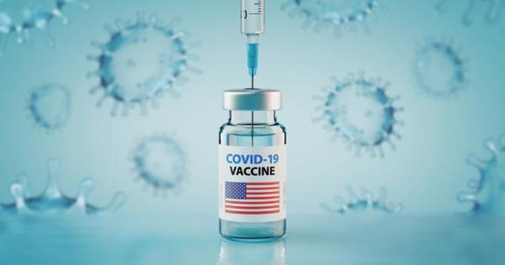
The jab changes biochemistry of Glial Cells
COVID-19 Vaccine Alters Biochemical Composition of Glial Cells
Janelle Blankenship, PharmD, for Medscape
March 10, 2022
The study covered in this summary was published on bioRxiv.org as a preprint and has not yet been peer reviewed.
Key Takeaways
Incubation of normal and tumor glial cells with the Pfizer-BioNTech COVID-19 mRNA vaccine (BNT162b2) led to alterations in the biochemical profile of various cell organelles.
The vaccine was shown to decrease concentrations of the oxidized form of cytochrome c in mitochondria, which decreases effectiveness of oxidative phosphorylation, reduces apoptosis, and decreases ATP production.
The investigators also observed alterations in lipid composition, decreases in amide I concentration, and histone modifications in the cell nucleus.
The biochemical changes were similar for normal astrocytes, astrocytoma (representing a mildly aggressive brain tumor), and glioblastoma (representing a highly aggressive brain tumor).
Why This Matters
This study investigated possible effects of the vaccine on the central nervous system.
The results of this study suggest that immune responses are reprogrammed by exposure to the vaccine.
Cytochrome c in the mitochondria of cells plays a key role in oxidative phosphorylation and apoptosis; therefore, the protein also plays a key role in cancer development. The authors indicate a potential association between cytochrome c, the immune system, and cancer development.
The investigators were able to demonstrate the utility of Raman spectroscopy and imaging for noninvasive monitoring of the biochemical profile in specific organelles. This methodology may potentially be used to contribute to further understanding of metabolic pathways, immune response, and cancer development.
Study Design
In this study, the investigators used normal human astrocytes (Clonetics NHA), human astrocytoma CCF-STTG1 (ATTC CRL-1718), and human glioblastoma cell line U87-MG (ATCC HTB-14). These cells were seeded on a CaF2 window in pure culture medium for 24 hours before they were supplemented with different vaccine doses (1 µL/mL or 60 µL/mL) and for different time periods (1 hour, 24 hours, or 96 hours).
After incubation with the vaccine, the biochemical alterations in various cell organelles were monitored using Raman imaging and spectroscopy.
P values ≤.05 were considered statistically significant.
Key Results
The concentration of the oxidized form of cytochrome c in the mitochondria, as determined by the normalized Raman intensity of the band at 1584 cm-1, decreased after incubation with the vaccine compared with the control samples. This decrease in cytochrome c concentration was statistically significant for the astrocytoma CRL1718 sample.
The investigators suggest that downregulation of cytochrome c after exposure to the vaccine may alter innate immune responses.
After incubation with the vaccine, statistically significant decreases in cytochrome c signaling were observed in lipid droplets and rough endoplasmic reticulum for all cells, all incubation times, and all vaccine doses.
Decreases in the intensity of the band at 1654 cm-1, which corresponds to amide I vibrations, were observed for the glioblastoma cells that were exposed to vaccine. This decrease indicates a change in the mitochondrial membrane potential and suggests a decline in adenine nucleotide translocator function.
Incubation with the vaccine was also associated with alterations in the nucleus caused by posttranslational modifications of histones.
Limitations
The authors did not state any limitations.
Disclosures
This work was supported by statutory activity 2021: 501/3-34-1-1.
The authors declared no conflict of interest.
The authors stated that the funders were not involved in designing the study, performing the study, or writing the manuscript.
This is a summary of a preprint research study, "Decoding COVID-19 mRNA Vaccine Immunometabolism in Central Nervous System: Human Brain Normal Glial and Glioma Cells by Raman Imaging," written by Halina Abramczyk from Lodz University of Technology and colleagues on bioRxiv.org provided to you by Medscape. This study has not yet been peer reviewed. The full text of the study can be found on bioRxiv.org.
For more news, follow Medscape on Facebook, Twitter, Instagram, and YouTube.

