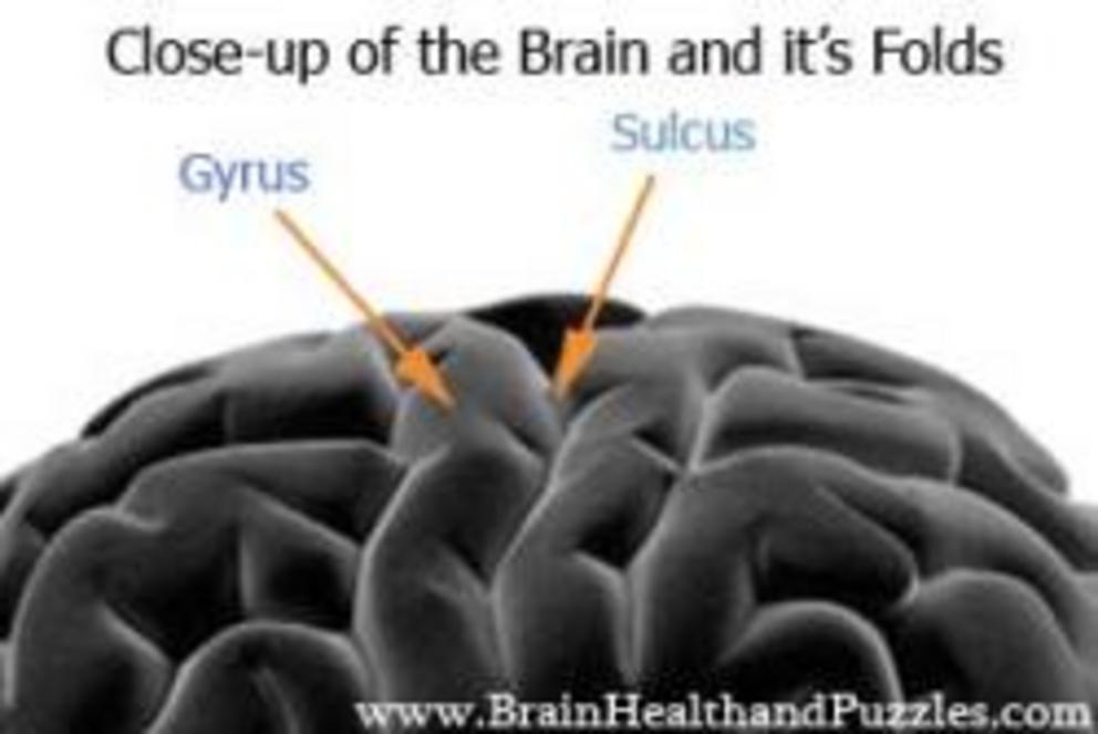
Abnormal brain folds in autism
... another clue from the gut?
The pieces of the autism puzzle have been in front of us for years:
-brain inflammation/larger brains in some, not all
-loss of language/abnormal use of language
-social thinking challenges
-sensory issues
-anxiety
-food allergies
-immune dysfunction/autoimmunity
-seizures
-gastrointestinal disorders/disease - reflux, colitis, inflammation
- constipation and/or diarrhea
-perseveration/obsessions and compulsions
-tics
Two new studies recently came out but are kind of a repeat of some past research but can be added in as clues. To aid in understanding, Gyrification means “the process of forming the characteristic folds of the cerebral cortex in the brain”. :
- A Longitudinal Study of Local Gyrification Index in Young Boys With Autism Spectrum Disorder :Local gyrification index (LGI), a metric quantifying cortical folding, was evaluated in 105 boys with autism spectrum disorder (ASD) and 49 typically developing (TD) boys at 3 and 5 years-of-age...At 3 years-of-age, this subgroup exhibited increased LGI ...relative to TD boys ...In summary, this study identified alterations in the pattern and development of LGI during early childhood in ASD. Distinct patterns of alterations in subgroups of boys with ASD suggests that multiple neurophenotypes exist and boys with ASD and disproportionate megalencephaly should be evaluated separately.
- Local Cortical Gyrification is Increased in Children With Autism Spectrum Disorders, but Decreases Rapidly in Adolescents : Extensive MRI evidence indicates early brain overgrowth in autism spectrum disorders (ASDs). Local gyrification may reflect the distribution and timing of aberrant cortical expansion in ASDs. ...Altered gyrification may reflect unique information about the trajectory of cortical development in ASDs.
So, aberrant cortical expansion basically means that there is an overgrowth of FOLDING in the brain. Scientific American put it this way: “Young people with autism show a lot of folding in patches of the left temporal and parietal lobes—regions responsible for processing sound and spatial information respectively, the study found. They also show excessive folding in temporal and frontal regions on the right side of the brain, including areas that govern decision-making and motor skills...They have less folding in one spot on the occipital cortex, the brain’s visual hub.”
As I said, this is not new information as other studies have reported the same -- a sampling:
- Paris, 12 January 2016 Is autism hiding in a fold of the brain ?
- 2014 - Atypical sulcal anatomy in young children with autism spectrum disorder
- 2011 - Early Brain Overgrowth in Autism Associated with an Increase in Cortical Surface Area Before Age 2
- 2008 - 3D Cerebral Cortical Morphometry in Autism: Increased Folding in Children and Adolescents in Frontal, Parietal, and Temporal Lobes
Most of this research claims -- an array of genetic changes and environmental conditions -- are responsible for this unusual folding.
Other research is overlapping here and the emphasis is again --- on the gut microbiome, a favorite topic of mine.
What they have done is look at bacteria, the type and the amounts, and then correlated those with the structure of the brain. This was an IBS (Irritable Bowel Syndrome) study, which has connections to autism - “an increased ratio between the phyla Firmicutes and Bacteroidetes (F-B ratio) which has been reported by several investigators [7–10]....Higher alpha diversity has previously been reported in an IBS subgroup [10], in patients with celiac disease [51], and with autism spectrum disorder [52], all syndromes which are often accompanied by IBS-like symptoms.
It is fascinating as the connections from bacteria types and amounts, to brain volume or folds has yet to be done for autism but a study would be very important.
Moderate-sized correlations with brain structure were observed for certain Firmicutes- and Bacteroides-associated taxa. For example, the Firmicutes-associated Clostridia (higher in IBS1) and the Bacteroidetes-associated Bacteroidia (lower in IBS1) showed correlations with the volume of several subcortical brain regions involved in sensory integration and modulation and the motor cortex. For the majority of these regions, increased volumes were observed with decreases in Bacteroidia taxa and increases in the Clostridia taxa...For the sensory brain regions, it is conceivable that neuroactive or proinflammatory metabolites generated by altered gut microbiota reach the brain, inducing neuroplastic changes. As most patients with IBS symptoms have a longstanding history of symptoms, often dating back to childhood, it is likely that such altered gut microbiota to brain signaling could have shaped the brain from early on in life.
Consider this too, from that same study -- In support of this possibility, we found that the surface area of the posterior insula was associated with the predicted abundance of 20 bacterial genes increased in the IBS1 group.
That’s right. BACTERIAL genes.
The identified genes included two that influence synthesis/degradation of GHB and glutamate. GHB is a neurotransmitter found naturally at high levels in the intestine that inhibits intestinal peristalsis via GABAB receptors and has sedative effects in the CNS [70, 71]. Glutamate is an excitatory neurotransmitter in the enteric nervous system and in the brain where it also plays an important role in synaptic plasticity….
Interestingly, this study may again show that we need to examine autism in this manner - bacteria vs the brain, especially the issue of abnormal folding:
Folding of the brain has been shown to be connected to intelligence . Could we find out more by looking at these connections of the “convoluted” folds and the gut bacteria? We could and hopefully it will be done ----
The low functioning autism group had a prominent shape abnormality centered on the pars opercularis of the inferior frontal gyrus that was associated with a sulcal depth difference in the anterior insula and frontal operculum. The high-functioning autism group had bilateral shape abnormalities similar to the low-functioning group, but smaller in size and centered more posteriorly, in and near the parietal operculum and ventral postcentral gyrus. Individuals with Asperger’s syndrome had bilateral abnormalities in the intraparietal sulcus that correlated with age, intelligence quotient, and Autism Diagnostic Interview-Revised social and repetitive behavior scores……
Early life perturbations of the developing gut microbiota can impact neurodevelopment and potentially lead to adverse mental health outcomes later in life ….What is clear is that this has profound
implications for our understanding of how environmental
exposures at specific stages of the lifespan may impact
neural plasticity to modify neurobehavioral development,
and what implications this may have for the risk of neurodevelopmental
and neuropsychiatric disorders.
What this could mean is that certain bacteria may actually change the architecture of the brain and functioning, explaining the spectrum of autism from the high functioning patients, to those moderately impacted, to those who are the most severely affected. Environmental exposures may have indeed caused the microbiome to become dysfunctional.
The Michael J. Fox Foundation has made progress in this line of research. Hopefully autism will follow--
….clumped proteins produced by bacteria in the gut cause brain proteins to misfold via a mechanism called cross-seeding, leading to the deposition of aggregated brain proteins. He also proposed that amyloid proteins produced by the microbiota cause priming of immune cells in the gut, resulting in enhanced inflammation in the brain. The research, which was supported by The Michael J. Fox Foundation…..

