Breakthrough in cell imaging could have major impact in crime labs
A Virginia Commonwealth University researcher has developed a procedure for identifying the source of cells present in a forensic biological sample that could change how cell types are identified in samples across numerous industries.
Many traditional techniques for distinguishing between saliva, blood, skin or vaginal tissue in an evidence sample are based on microchemical reactions that can be prone to false-positive or false-negative results, according to the researcher, Christopher Ehrhardt, Ph.D., an associate professor in the Department of Forensic Science in the College of Humanities and Sciences. Additionally, they may be difficult to use on aged or heavily degraded samples.
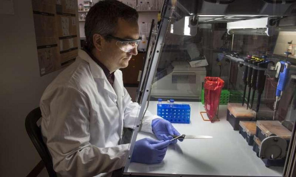
Unlike traditional forensic testing methods, Christopher Ehrhardt’s procedure can be used to identify different cell types in a sample without damaging the sample. Credit: Kevin Morley, University Relations
"The information is often limited," Ehrhardt said. "And when using conventional methods, you have to be prepared to consume part of the sample in most cases, which decreases the value of it."
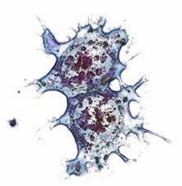
Ehrhardt's procedure aims to change that. He begins by taking microscopic images of the individual cells using a benchtop microscope or a flow cytometer—a device used in cell biology that photographs individual cells encased within drops of water. Ehrhardt then makes measurements that capture size, shape and fluorescent properties of the cells. Those measurements are then analyzed using machine learning algorithms—in this case computer software programmed to recognize characteristics of the images—to correlate them with cell type.
"This new procedure can be used to identify different cell types in a sample as well as potentially indicate some attributes of the individuals who deposited the cells, like age, sex and so forth," Ehrhardt said. "And the best part is that the procedure is nondestructive. After imaging, the cells can be used to generate a DNA profile. This is really important since many samples are very little biological material, so the more information you can get without consuming the sample, the better."
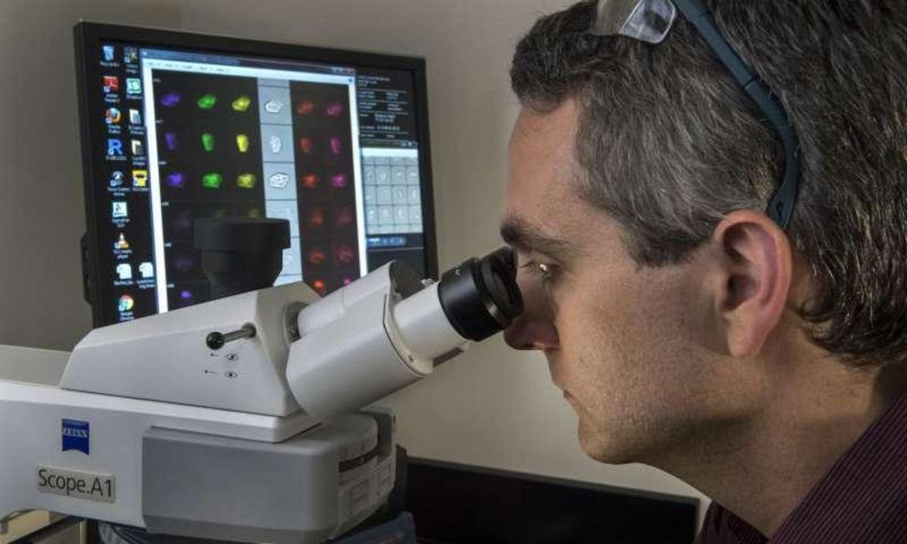
Ehrhardt's process begins by taking microscopic images of the individual cells using a benchtop microscope or a flow cytometer — a device used in cell biology that photographs individual cells encased within drops of water. Credit: Kevin Morley, University Relations
Brent Fagg, technology manager with VCU Innovation Gateway, said forensic laboratories could use this new procedure to improve the efficiency of their testing.
"Traditional forensic testing methods are time-consuming, destructive to samples, and unable to determine the abundance of cell types in a sample," he said. "Using our new procedure, labs will be able to analyze aged or degraded samples in a quick and nondestructive manner—and with much better results."
Fagg said forensic analysis is just one possible application for this new procedure. It also could be used in areas such as pharmaceutical and health care, and even to monitor exposure to disease.
"There are a number of industries that could benefit from this new cell type identification procedure," he said. "And adopting this technique couldn't be easier, as it uses lab equipment common in biology laboratories."
The procedure was described in a study, "Rapid differentiation of epithelial cell types in aged biological samples using autofluorescence and morphological signatures," that was published May 18 in the journal PLOS One.